现在,3D打印技术已经越来越多的在各领域得到了应用,日前,内蒙古自治区肿瘤医院微创介入中心利用国际先进的数字化医疗3D打印模板导向技术,为一名上颌窦癌患者成功实施了放射性粒子植入术即组织间放疗。
患者是一名年轻女性,失去了手术机会,在外放疗及化疗效果不佳的情况下来到内蒙古自治区肿瘤医院就诊。为保护患者五官功能及容貌,医院微创介入中心采用了疗效确切的放射性粒子植入方法治疗。该方法是将带有放射性的粒子植入肿瘤内,对肿瘤照射,由于粒子有效照射距离短,只有1.7cm,既可很好的杀伤肿瘤,又能保护正常组织和器官,是目前治疗恶性肿瘤有效办法之一。
据冯铁虹教授介绍,既往放射性粒子植入术是在CT或超声引导下穿刺植入,因骨骼、血管、神经及器官等原因,植入的粒子往往不能完全包括肿瘤,达不到理想的治疗效果,致使二次手术植入粒子或肿瘤缩小后根据情况再次补充植入粒子治疗。为了准确进针位置和深度,并且避开骨骼、血管、神经等,在手术治疗时需要对患者部位进行多次CT扫描定位,增加了患者不必要的损伤和医疗费用。而数字化医疗3D打印模板导向技术弥补了上述不足。
数字化医疗3D打印模板导向技术首先是利用CT扫描后三维立体重建数据,在计算机软件中模拟进行对病变组织穿刺。然后利用3D打印技术根据病变组织体表形状打印出3D适型模具,通过计算机提供的模板上的每一个穿刺通道,将穿刺针送入病变组织。
冯铁虹教授的研究小组将CT扫描后得出的数据送到软件开发单位之一的北京工业大学进行3D适型模具的制作。在手术中,医生将3D适型模具放置在患者面部,利用数字化设备和3D适型模具对患者进行穿刺。相比以前单纯利用数字化设备进行CT或超声引导下穿刺植入,准确性大大提高了。“以往没有利用3D技术的时候,计算机可以说只能做一个大致的手术规划。在手术过程中,为了保证进针准确,避开其他器官,我们甚至需要做四五次甚至十几次CT扫描。现在有了3D打印的模板,进针定位、深度一切都提前设定好了,粒子植入更加精准,使治疗效果大大提高了。而CT扫描也仅仅只做了两次。”冯铁虹教授说。
据冯教授介绍,单纯利用数字化设备的同样的手术往往需要近两个小时的时间,而引入3D打印后这个手术只用了半个小时就完成了。而不仅如此,该技术还简化了手术程序,使放射性粒子植入治疗肿瘤的手术更利于在基层医院普及推广 。
目前在国内外,已有将3D打印技术用在骨科临床领域,而此次将3D打印技术用在放射性粒子植入术中尚为首次,是临床治疗的一次新的突破。
Today, we learn about yet another successful use of 3D printed models within the medical field. This story comes out of China, specifically the Inner Mongolia Autonomous Region, where Professor Feng Tiehong has utilized a 3D printed template model in order to help destroy cancer in a young woman. This woman (remains unnamed) was suffering from cancer of her maxillary sinus area, and typical surgery to remove the legions would have left her with major scaring and disfiguring of her face. However, thanks to a technology called radioactive seed implantation, along with 3D printing, surgeons were able to more efficiently destroy the cancer cells.
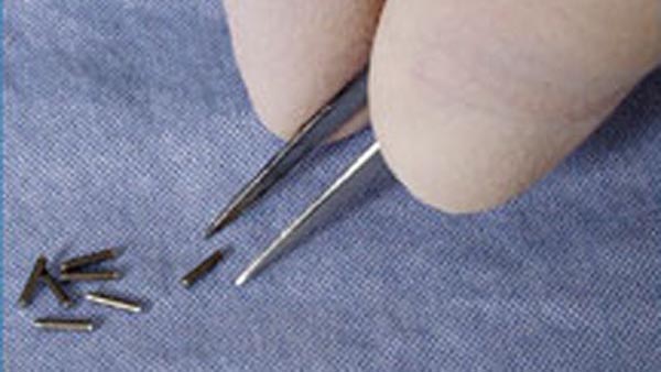
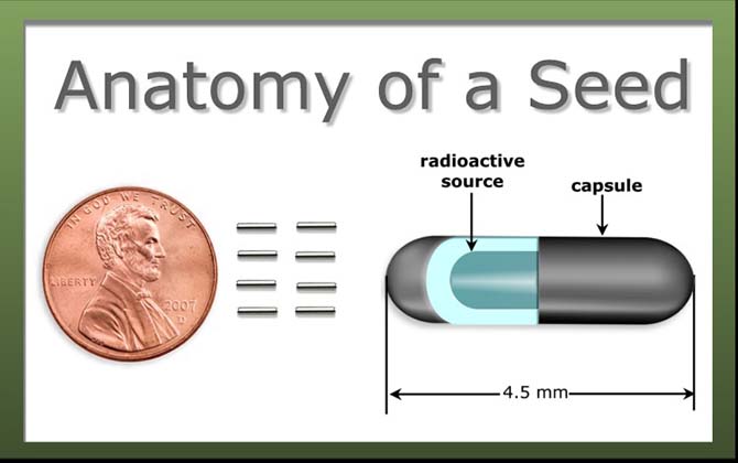
Radioactive seed implantation has been used for years. It is a process by which radioactive particles (usually titanium) are injected into cancer tissue within a person’s body. It is most commonly used for instances of prostate cancer, but can be used for other types of cancers as well. These “seeds” are usually smaller than a grain of rice. When injected into the tissue, they provide radiation to the surrounding cells, within a 1.7 cm radius. Cancer cells are much more sensitive to the radiation, thus surrounding tissue is not effected, while the cancer cells are killed.
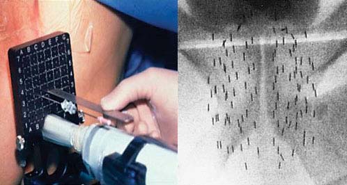
Typically doctors use many CT scans, as well as ultrasound, in order to determine where to best inject the radioactive seeds. However, this process is a long and rather inaccurate one, requiring many attempts and many additional scans in order to get the seeds put in just the right places. Professor Fen Tiehong, however, has introduced a method in which a 3D printed template is created, where the injections can precisely be placed in the exact locations.
“In the past there has been no use of 3D technology, and the computer can be said to only do a general surgical planning,” says Professor Gen Tiehong. “In the past, during the procedure, in order to ensure the accuracy of the needles, and avoid other organs, we even need to do four or five or even ten separate CT scans. Now With 3D printing templates, needle positioning, depth, and everything else is set well in advance. This makes seed implantation more precise, so that treatment is greatly improved, while CT scans are only needed twice.”
In what is referred to as “digital medical 3D printing template oriented technology”, doctors were able to create a model of this woman’s sinus area, using CT scan data and computer software to simulate the diseased tissue. Then a 3D conformal mold was printed out according to the shape of the cancer tissue’s surface. The template provided a puncture channel, created by computer software, showing and guiding doctors exactly where to place the radioactive seed-bearing needles. This template was placed on the woman’s facial area, and the needles were inserted into the channels in order to precisely place each individual radioactive seed.
Tiehong says that this technology has allowed surgery time to be cut from two hours down to only 30 minutes. He hopes and believes that 3D printing will gain much more mainstream traction within medical practice, and thinks the technology could give individuals an option for surgery that was not previously available.
“In the past, because of the difficulty of the radio active seed surgery, many hospitals rarely carried it out, but now 3D printing, greatly reduces the difficulty of the surgery,” explains Tiehong.